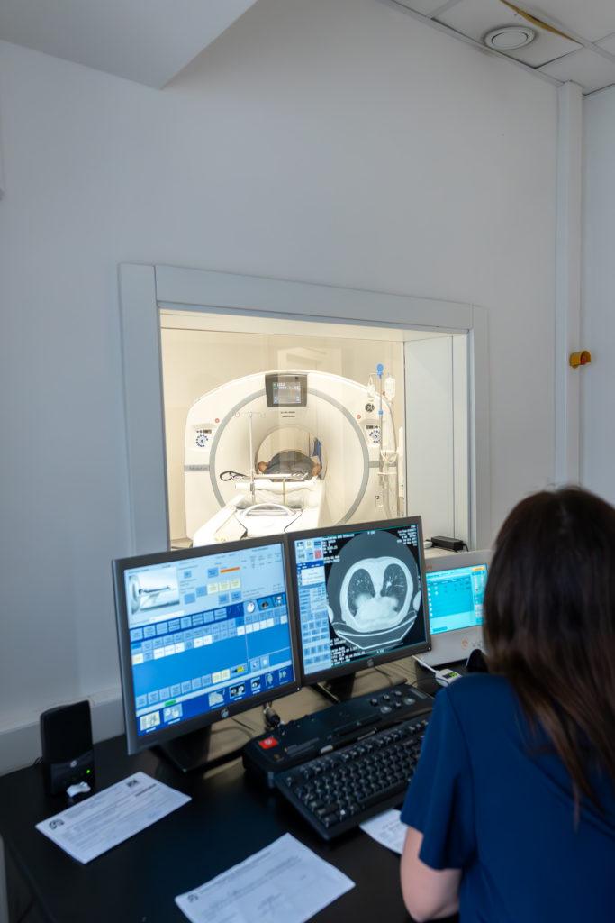
Department of Radiological Diagnostics
The department is staffed, organized, technically equipped, and spatially adapted to fulfill its primary purpose within the Institute, which is to perform various examinations in the field of conventional X-ray, ultrasound, and CT diagnostics.
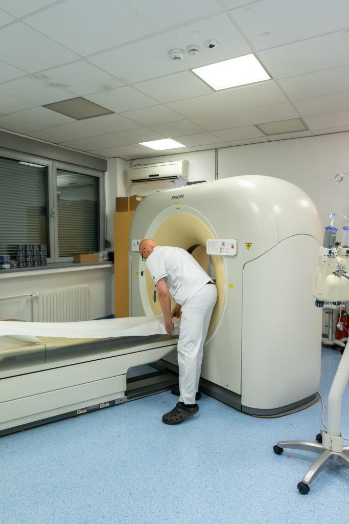
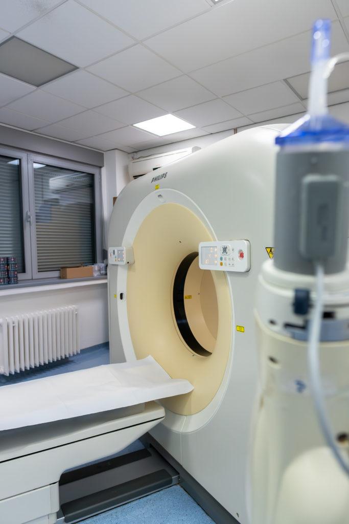
Patients are examined in six rooms for conventional radiography and radioscopy, one for ultrasound diagnostics, and one for computerized tomography (CT scanner).
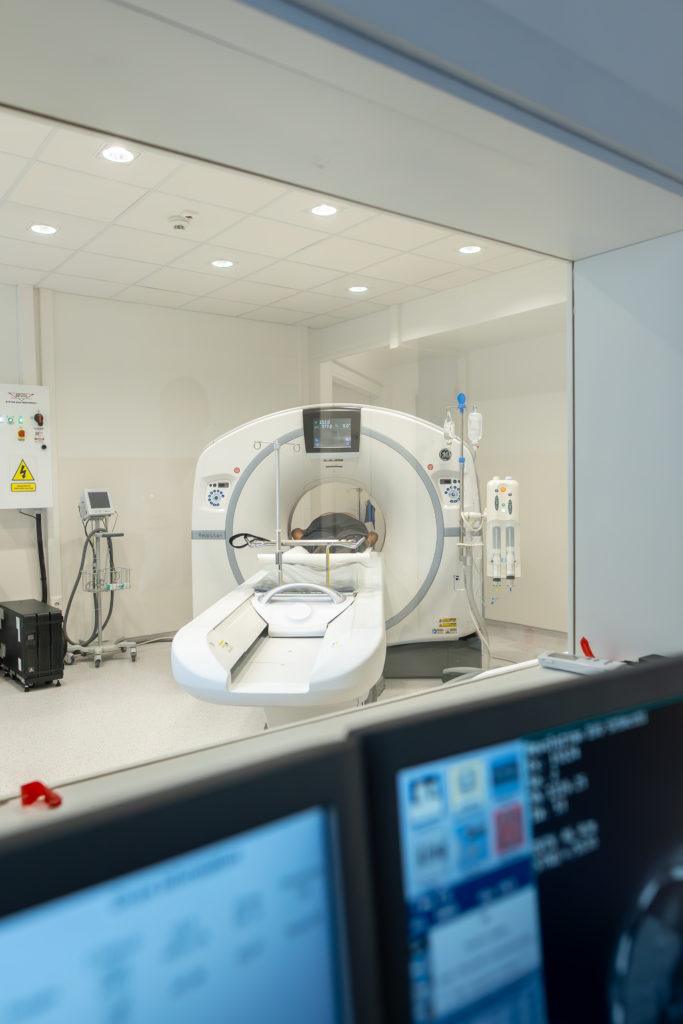
In the last five years, patients in the computed tomography (CT) office have been examined on the GE LIGHTSPEED 64 slice multi-slice CT device, which allows for scanning the entire volume of the thorax during a single breath hold and creating a three-dimensional, high-resolution image without artifacts. Transthoracic punctures under CT guidance are also performed here.
Transthoracic puncture is a minimally invasive diagnostic method that provides a cytological tissue sample for examination to clarify the etiology of observed changes in the lungs, pleural space, or chest wall. The sample obtained from the puncture is cytologically analyzed for the purpose of establishing an etiological diagnosis. Our institution performs approximately 250 such examinations annually.
The use of the MEDRAD automatic injector enables the efficient execution of all CT angiographic procedures, including CT pulmonary angiography.
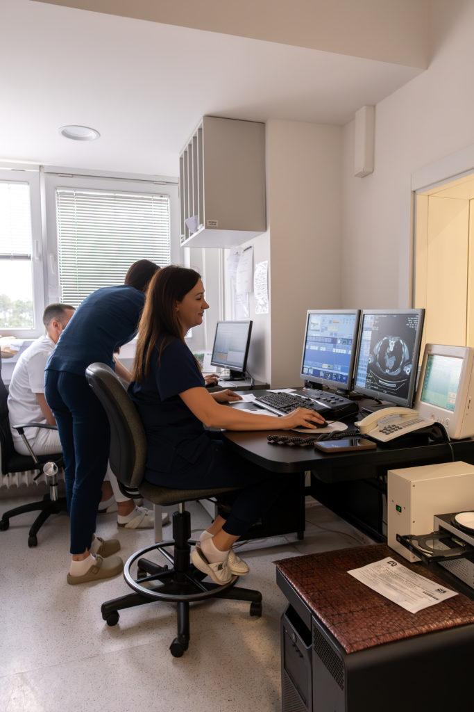
The review of obtained images is performed on the GE workstation Advantage 4.4, where various software packages are applied, in addition to the usual (native and post-contrast standard and targeted axial examinations), high-resolution lung examinations (HRCT), 3D, MPR, and virtually endoscopically reconstructed examination techniques, volumetry of pulmonary lesions, as well as CT angiography and CT coronary angiography.
The department for conventional radiography and radioscopy is equipped with:
A digital X-ray system GE DEFINIUM 8000, which is among the most advanced radiographic systems with a flat panel detector. Automatic exposure control allows for the interruption of acquisition in a thousandth of a second, further contributing to the reduction of patient exposure to X-ray radiation.
The device is also intended for radiographic subtraction of bone structures – DUAL ENERGY advanced method that allows for the separate display of soft tissues and bone structures that obscure soft tissues in standard images.
The Institute utilizes two mobile radiography machines, located at the Clinic for Thoracic Surgery and in the Intensive Care Unit. The radiographs produced there are then developed in a digital imaging device.
Since August 2014, the Service has upgraded the ultrasound room with a new GE LOGIQ F8 ultrasound machine, which allows us to obtain images of optimal resolution for the daily examination of a large number of patients. The majority of examinations include abdominal ultrasound, as well as the examination of pleural spaces, with localization and evaluation of pleural effusions.
The Service has the necessary personnel and technical prerequisites, and the legal framework (Law on Electronic Signatures) has also been established for conducting teleradiology consultations. By establishing a teleradiology connection through high-speed internet connections, the reporting time is reduced, quality is guaranteed, and it allows for bridging the shortage of personnel for radiological diagnostics.
The Radiological Diagnostics Department of IPBV employs four specialist radiologists, four residents in radiology, and 16 senior radiological technicians who, along with other staff, perform up to 80,000 individual patient examinations annually (72,000 X-rays, 6,000 CT scans, and 3,500 ultrasound examinations). The number of examinations has a tendency to continuously increase.
Throughout the year, employees enhance their skills through lecture programs, seminars, and practical training. The Radiological Diagnostics Department actively participates in conducting clinical and institutional studies and other projects. Recent improvements in operations have also been achieved with the newly implemented hospital information system.
It is of utmost importance to enhance radiation protection measures, both for patients and for all staff.